Lv Non Compaction Echo
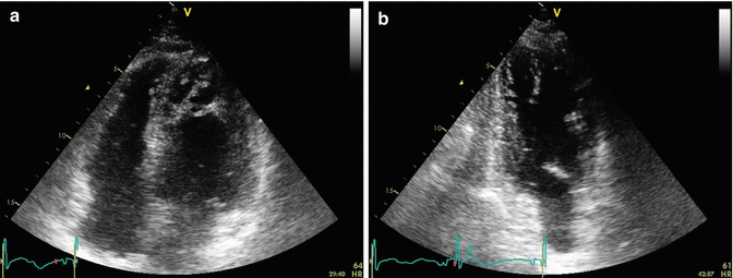
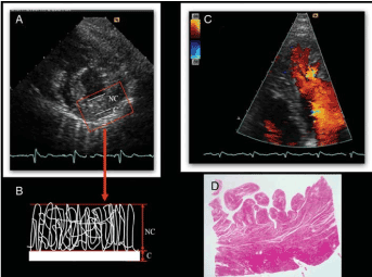
Left Ventricular Noncompaction Cardiomyopathy Panel A Panel C Panel D Panel B LV RV LV RV LV LV T h r o m b o e m b o l i c c o clot clot LV RV MRI • This is the major and primary diagnostic imaging criterion • It is critical to obtain images that are not foreshortened and are perpendicular to the ventricular long-axis view Tips & Tricks in the Echo Lab.
Lv non compaction echo. It can be associated with left ventricular dilation or hypertrophy, systolic or diastolic dysfunction, or both, or various forms of congenital heart disease. In left ventricular non-compaction cardiomyopathy (LVNC) the lower left chamber of the heart, called the left ventricle, contains bundles or pieces of muscle that extend into the chamber. During development, the heart muscle is a sponge-like network of muscle fibers.
The prevalence of left ventricular non-compaction is not well established. By itself, the diagnosis of LVNC does not coincide with that of a “cardiomyopathy” because it can be observed in healthy subjects with normal LV size and function, and it can be acquired and is. Echoes from 100 patients, of which 51 had received the diagnosis of LV non-compaction (NC), were reviewed.
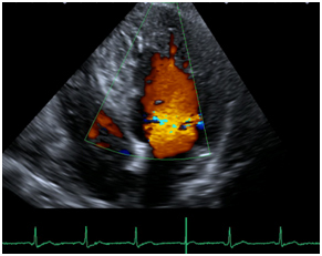
Transthoracic echocardiography is also useful for detecting associated lesions like muscular ventricular septal defects or supra mitral ring along with non-compaction. LVNC can sometimes be caused by mutations in genes that encode for structural proteins of heart muscle. These are best visualized on color flow Doppler of the left ventricle using apical windows.
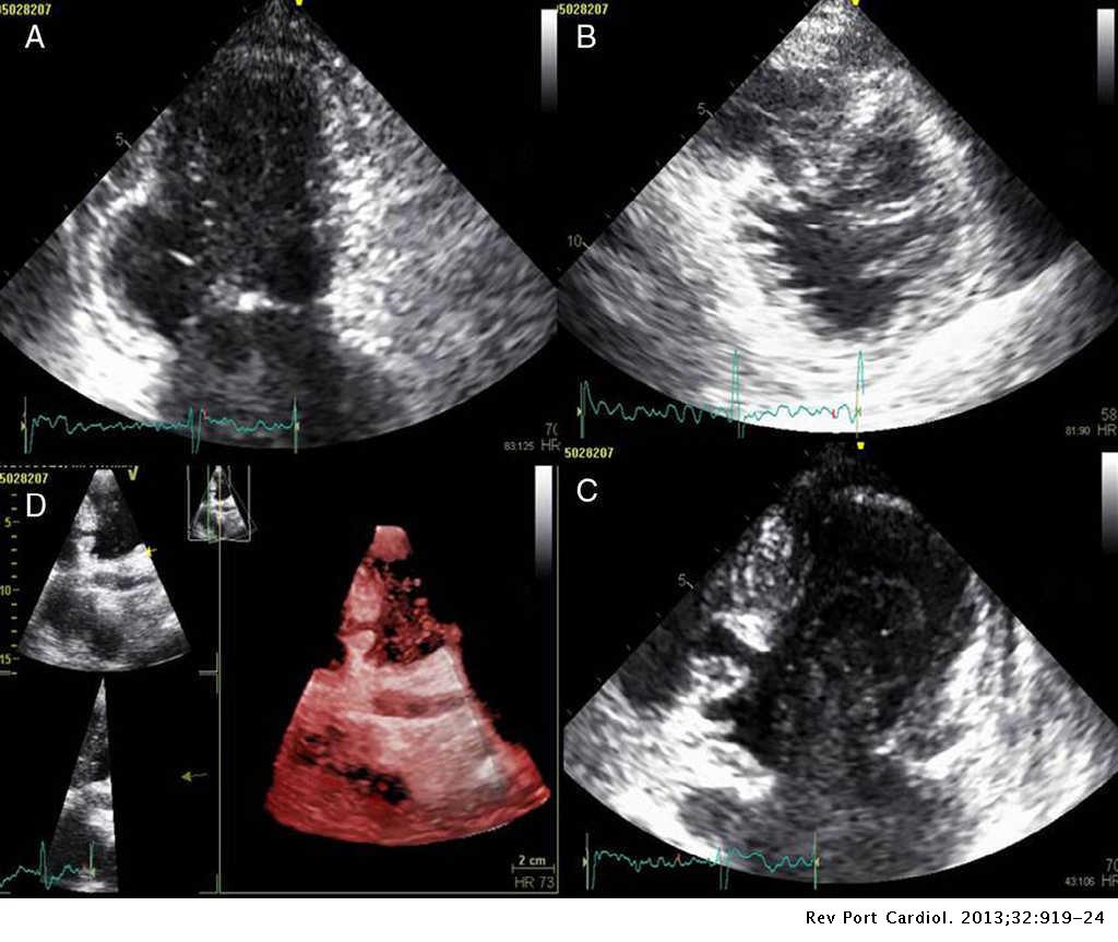
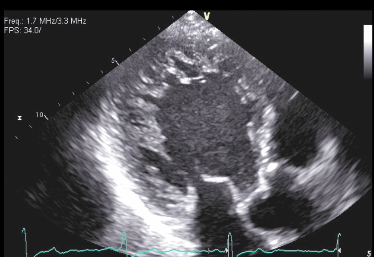
LV noncompaction is characterized by numerous prominent trabeculations and deep intertrabecular recesses in communication with the ventricular cavity. A new cardiomyopathy is presented to the clinician". Non-compaction cardiomyopathy (NCM) is a myocardial disorder, which is thought to occur due to the failure of left ventricle (LV) compaction during embryogenesis, leading to distinct morphological characteristics in the ventricular chamber.1 It was first described about 80 years ago, in association with complex congenital heart diseases.
Moderate to severely depressed left ventricular systolic function. Using 2-D echocardiography, less than 1% of patients are diagnosed with this entity. Heavily trabeculated left ventricle, most pronounced in the apical and left ventricular free wall.
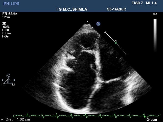
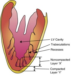
1,2 The overall prevalence is unknown, but a review of patients having. The ratio of noncompacted to compacted myocardium here is roughly 2:1. "Noncompaction of the left ventricle:.
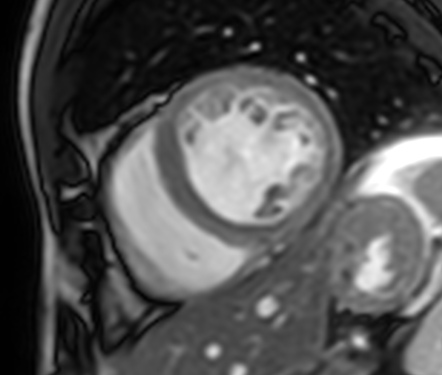
Individual variability is extreme, and trabeculae represent a sort of individual “cardioprinting.”. The value of cardiac magnetic resonance imaging in the diagnosis of isolated non-compaction of the left ventricle. It affects approximately 1 in 700 people.
10.1093/eurheartj/ehp595 Crossref Medline Google Scholar. This variance may be related to the difference in spatial resolution between these 2 imaging techniques. MedlinePlus - Health Information from the National Library.
Prominent left ventric- ular (LV) trabeculae, deep intertrabecular recesses, and the thin compacted layer (1).The spectrum of morphologic variability is extreme, ranging from hearts with a nearly absent compacted layer and an almost exclusively trabecular compo- nent in the LV apex, to hearts with prominent trabeculae and deep alternating recesses, but a well-represented compacted layer. It is characterized by trabeculated myocardium with adjacent deep intertrabecular recesses communicating with the LV cavity 1. Vatta M, Mohapatra B, Jimenez S, et al.
LVNC is a condition of the heart where the walls of the left ventricle (the bottom chamber of the left side of the heart) are non-compacted. "Non-compaction of the Left Ventricular Myocardium - From Clinical Observation to the Discovery of a New Disease". Left ventricular noncompaction (LVNC) cardiomyopathy is a new and as yet unclassified cardiomyopathy with an estimated prevalence of 0.014% to 0.17%.
In LVNC the inside wall of the heart is spongy or grooved, instead of smooth. Affected individuals are at risk of left or right. Signs and symptoms of LVNC vary, but may cause life-threatening abnormal heart rhythms and weakness of the heart muscle.
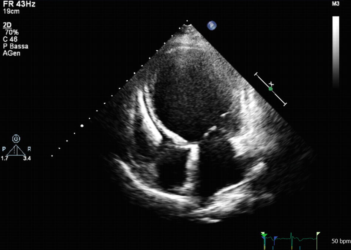
In non-compaction left ventricle, there are prominent trabeculations. Arrest of compaction process during normal heart development is thought to be the cause of LVNC 1,2.Echocardiographic demonstration of the apical trabeculation of left ventricle and blood flow from ventricular cavity into the intertrabecular recesses are the main pathologic. This study used speckle-tracking echocardiography to evaluate LV twist in patients with LVNC and determine whether abnormal LV twist is associated with more adverse LV remodelling.
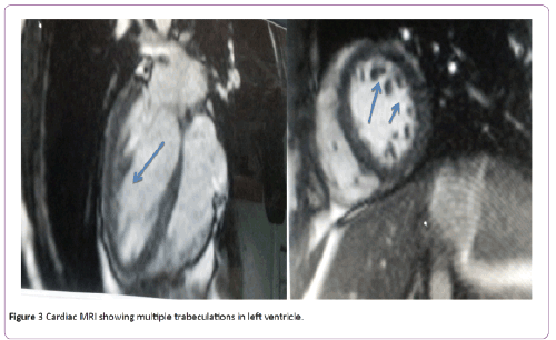
Left ventricular noncompaction 1. Sao Paulo Med J. Images of the left ventricle showed a 2-layer structure with a compacted, thin epicardial band and a much thicker noncompacted endocardial layer of trabecular meshwork.
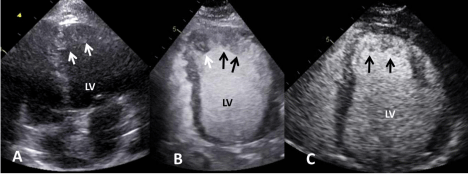
Hypertrabeculation of LV can be a benign finding but can also be associated with left ventricular non-compaction (LVNC), hypertrophic cardiomyopathy, dilated cardiomyopathy and heart failure. Measurement of trabeculated left ventricular mass using cardiac magnetic resonance imaging in the diagnosis of left ventricular non-compaction. Our echocardiography laboratory was consulted to determine whether a patient's echocardiogram would fulfill the criteria of left ventricular noncompaction cardiomyopathy (LVNC).
Raysa Morales-Demori, MD Left ventricular non compaction (LVNC) is a type of cardiomyopathy which is characterized by the presence of prominent trabeculations in the left ventricle with deep recesses between the trabeculations and a thin compacted myocardial layer. Left ventricular noncompaction (LVNC) is a relatively new entity. Left ventricular noncompaction is a rare unclassified cardiomyopathy with markedly prominent apical trabeculae with deep intertrabecular recesses (Fig.
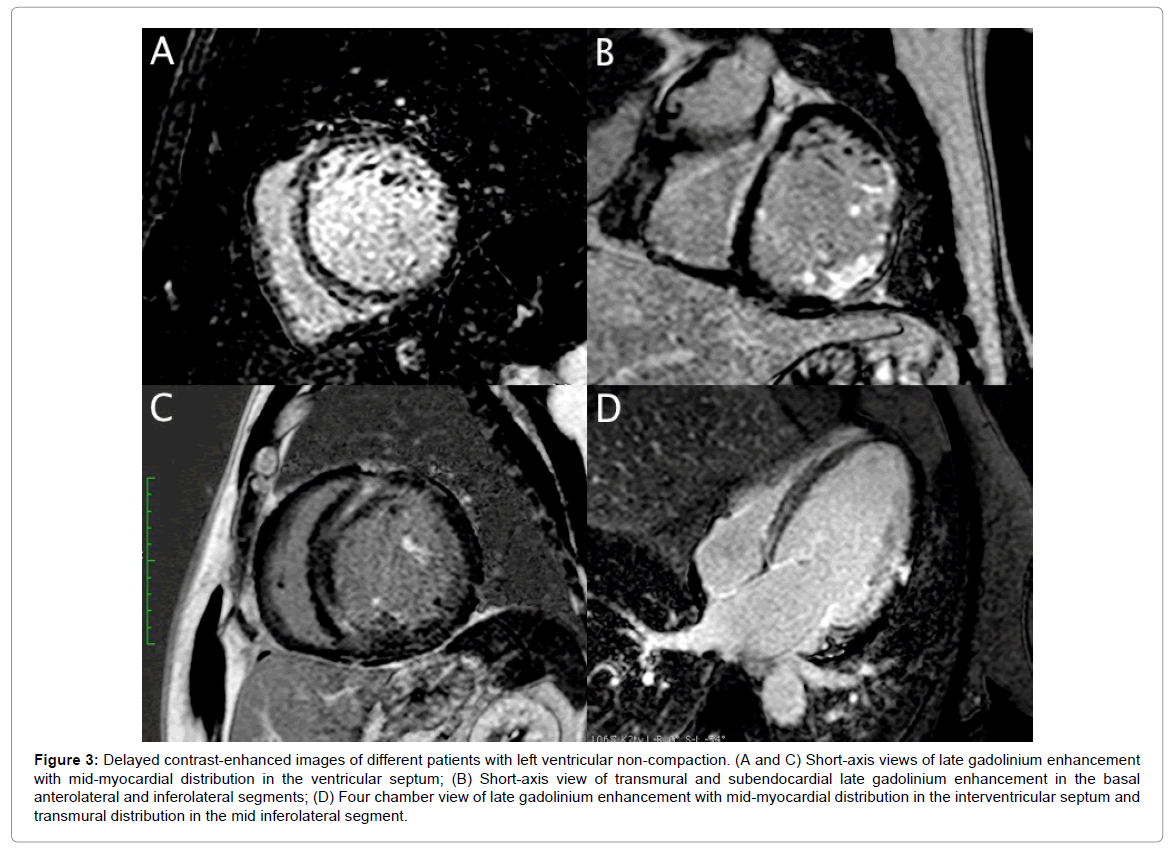
Left ventricular noncompaction is a rare cardiomyopathy that should always be considered as a possible diagnosis because of its potential complications. Superior contrast-to-noise ratio and signal-to-noise ratio Unlimited imaging planes Ability to use tissue characterization in the diagnosis High sensitivity and specificity Interobserver reproducibility of trabecular mass measurement in Jacquier criteria reported to be high Interobserver and intraobserver. Distinction between LV NC and non-specific dilated cardiomyopathies (DCMs) remains often challenging.
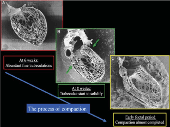
1 It is a rare primary cardiomyopathy that is an isolated finding in 74% of cases, or is associated with congenital heart disease in 26% of cases. Left ventricular noncompaction (LVNC) also known as ‘spongy myocardium’ is a rare abnor-mality of the left ventricular (LV) wall that results from intrauterine developmental arrest of the normal compaction process of the myocardium during the first trimester leading to the formation of two layers of the myocardium:. Left ventricular noncompaction (LVNC) is a rare heart condition.
Agreement between the three reviewers occurred in 65% of the cases. Due in part to improved imaging with echocardiography and magnetic resonance imaging, clinical awareness and appreciation for the marked heterogeneity of this disorder is increasing. Left ventricular noncompaction 2.
The adequacy of diagnosis of non-compaction depends very much on experience and knowledge of the investigator. We aimed to investigate the compaction process of the LV myocardium during the normal gestation period and provide reference for echocardiographic diagnosis of a fetus with ventricular myocardium noncompaction. Isolated left ventricular noncompaction (LVNC) is a genetic cardiomyopathy characterized by prominent ventricular trabeculations and deep intertrabecular recesses, or sinusoids, in communication with the left ventricular cavity.
We sought to find additive tools comparing the longitudinal strain characteristics …. E4 Rigopoulos A , Rizos IK , Aggeli C , et al. Echocardiography widely available Portable investigation Cost-effective test:.
Left ventricular non-compaction (LV NC) is characterized by abnormal trabeculations that are mainly at the LV apex. The thickness and ratio of noncompacted and compacted layers of the left ventricular (LV) myocardium in the normal fetus were investigated by fetal echocardiography. These pieces of muscles are called trabeculations.
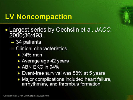
The low prevalence of patients with this cardiomyopathy presents a unique challenge for large, prospective trials to assess its pathogenesis, management, and outcomes. It is the third most frequent cardiomyopathy in children (~9%). Review of the 35 discordant cases resulted in agreement in 24 while 11 remained questionable.
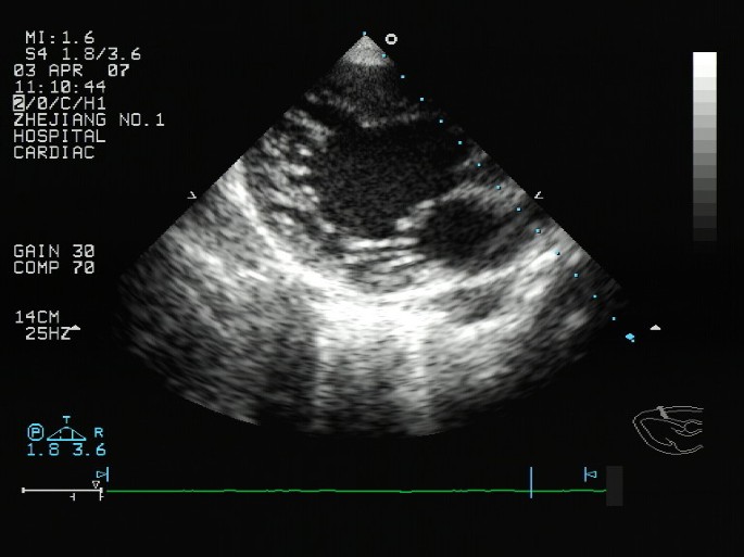
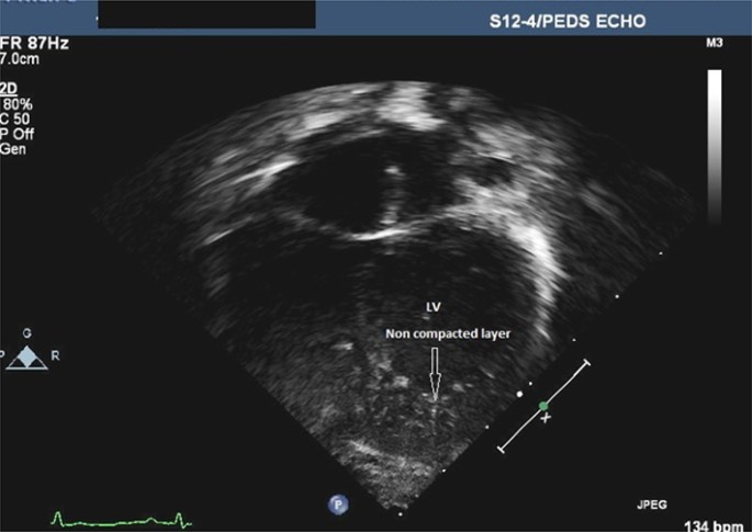
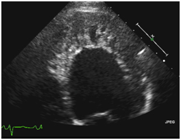
Closer to the apex the noncompacted myocardium is more evident with trabeculations seen in the anterolateral wall and inferior wall. Left ventricular noncompaction (LVNC) is a distinct phenotype characterized by prominent LV trabeculae and deep intertrabecular recesses 1,2. NVM is recently included in the 06 classsification of cardiomyopathies as a Genetic Cardiomyopathy 3.
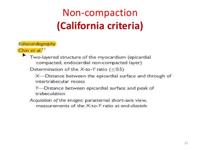
This gives the left ventricle a characteristic 'spongy' look (a bit like honeycomb). More recently, Chin et al 2 reported the isolated form. Microbiomes and Aging with Rob Knight - Research on Aging - Duration:.
De Groot-de Laat L., Krenning B., ten Cate F., et al.Usefulness of contrast echocardiography for diagnosis of left ventricular noncompaction Am J Cardiol, 95 (05), pp. Left ventricular non-compaction, the most recently classified form of cardiomyopathy, is characterised by abnormal trabeculations in the left ventricle, most frequently at the apex. Echocardiographic Criteria for LV Noncompaction The past two decades have witnessed significant advances in tissue harmonics and image resolution in echocardiography, which has enabled detailed assessment of the ventricular myocardium.
1990-First diagnostic criteria • LVNC X (distance between epicardial surface and trough of the intertrabecular recesses) Y (distance between epicardial surface and peak of the trabeculations) If X/Y< 0.5 if it progressively. Left ventricular non-compaction (LVNC) is a condition of the heart where the walls of the left ventricle (the bottom chamber of the left side of the heart) are non-compacted, causing channels to form in the heart muscle. This gives the left ventricle a 'spongy' look (a bit like honeycomb).
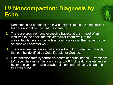
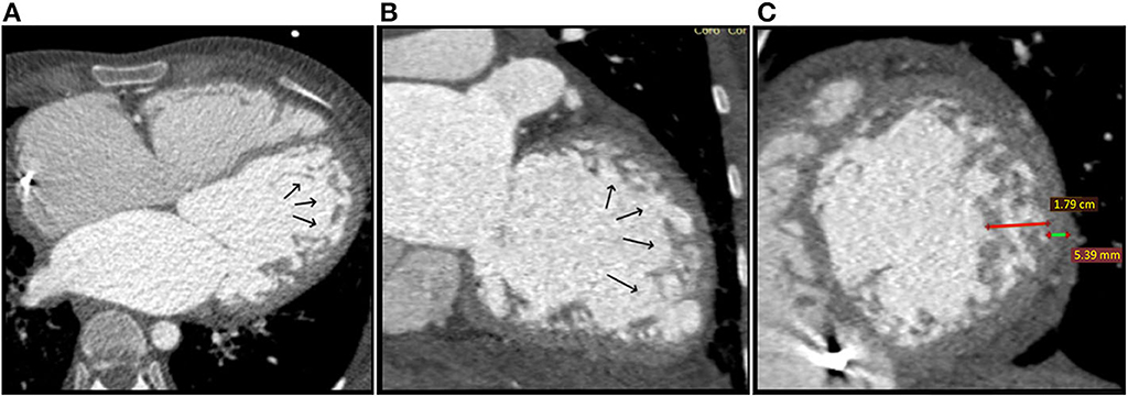
NCCM is characterized by excessive trabeculations typically involving the left ventricle (LV) with >2:1 ratio of noncompacted:compacted myocardium. Left ventricular (LV) hypertrabeculation is defined by the presence of three or more trabeculations apically and to the level of papillary muscles. To discuss diagnostic criteria for and the advantages and limitations of these imaging techniques;.
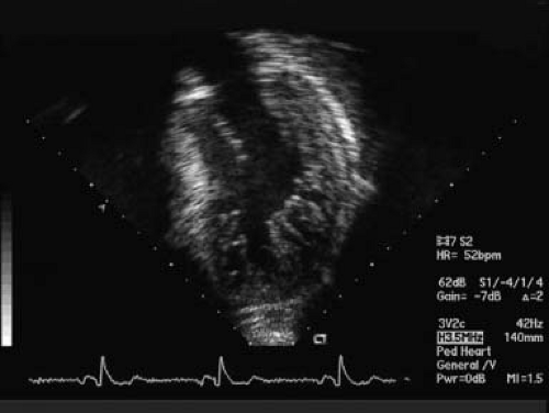
Apical 4 chamber view shows multiple trabeculations and deep recesses at the ventricular apex. Rarely, more than 3 prominent trabeculations that is the so-called LV noncompaction of ventricular myocardium (NVM) can be found at autopsy and by various imaging techniques including echocardiography and MRI etc. Eur Heart J.
Mutations in Cypher/ZASP in patients with dilated cardiomyopathy and left ventricular non-compaction. And to describe pitfalls that can lead to misinterpretation of findings of LVNC. Left ventricular (LV) twist is an important component of systolic function.
Echocardiography is the standard tool for diagnosis, and CMR is very useful to confirm or rule out this disease, especially when the apex is difficult to visualise. Using cardiac MRI, approximately 3% of patients have evidence of LV non-compaction. Isr Med Assoc J 09;11:313-4.
LVNC was previously also called spongy myocardium or hypertrabeculation syndrome but these terms should not be used interchangeably with LVNC 3. Non-compaction of the left ventricle, also known as spongiform cardiomyopathy or left ventricular non-compaction (LVNC) is a phenotype of hypertrophic ventricular trabeculations and deep interventricular recesses. The effect of abnormal LV twist on adverse remodelling of the heart in left ventricular noncompaction (LVNC) is unknown.
Eft ventricular noncompaction (LVNC) is de- fined by 3 markers:. Left ventricular noncompaction (LVNC) describes a ventricular wall anatomy characterized by prominent left ventricular (LV) trabeculae, a thin compacted layer, and deep intertrabecular recesses. Left ventricular noncompaction (LVNC) is a cardiomyopathy associated with sporadic or familial disease, the latter having an autosomal dominant mode o….
LV ejection fraction ranged from 4-%. Through ECHO window 3. The objectives of this article are to review the imaging findings of left ventricular noncompaction (LVNC) at echocardiography, cardiac MRI, and MDCT;.
Left ventricular non-compaction, also known as LVNC, spongy myocardium or hypertrabeculation syndrome, is a pathologic cardiac condition in which the myocytes exhibit a “spongy” appearance. Left ventricular thrombi in a patient with left ventricular non-compaction – visualisation of the rationale for anticoagulation. Isolated left ventricular noncompaction (LVNC), also called spongiform cardiomyopathy is a rare cause of heart failure.
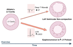
Noncompaction of the ventricular myocardium, also called left ventricular noncompaction (LVNC), is a rare congenital abnormality seen in only 0.05% of adults .It is characterized by spongy myocardium and results from arrest of the compaction of the loosely interwoven meshwork of myocardial fibers during endomyocardial morphogenesis between 5-8 weeks of fetal life. This causes channels to form in the heart muscle, called trabeculations. Coinciding with these developments has been an increasing number of reports of athletes with LVNC.
Left ventricular noncompaction (LVNC) is an heart disorder characterized by a heart that has not fully packed together its individual tissue layers, leading to deep recesses within the muscle wall.

Anzcmr Non Compaction Cardiomyopathy S Chen

Left Ventricular Non Compaction Youtube
Left Ventricular Noncompaction
Lv Non Compaction Echo のギャラリー

Fetal Noncompaction Cardiomyopathy And Histologic Diagnosis Of Spongy Myocardium Case Report And Review Of The Literature
Hypertrophic Cardiomyopathy Associated With Left Ventricular Noncompaction Cardiomyopathy And Coronary Fistulae A Case Report One Genotype Three Phenotypes Revista Portuguesa De Cardiologia English Edition
A Patient With Abnormalities Of The Coronary Arteries And Non Compaction Of The Left Ventricular Myocardium Resulting In Ischaemic Heart Disease Symptoms Dabek Folia Morphologica

Isolated Left Ventricular Noncompaction In Sub Saharan Africa A Clinical And Echocardiographic Perspective Circulation Cardiovascular Imaging
Left Ventricular Non Compaction Mid Myocardial Distribution Of Late Gadolinium Enhancement In Compacted Segments Omics International
Left Ventricular Noncompaction Cardiomyopathy Thoracic Key

Limitations In The Diagnosis Of Noncompaction Cardiomyopathy By Echocardiography
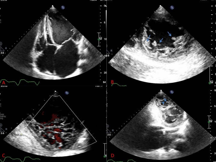
Multiple Thrombi In The Lv In Non Compaction Cardiomyopathy American College Of Cardiology
Multimedia Library Boston Children S Hospital

Clinical And Echocardiography Features Of Diagnosed In Adulthood Isolated Left Ventricular Noncompaction A Case Series Study Huang Wh Sung Kt Tsai Jp Lo Ci Hsiao Cc Kuo Jy Su Ch Chen Mr

Isolated Left Ventricular Noncompaction Cardiomyopathy A Transient Disease

Left Ventricular Noncompaction Diagnosed Following Graves Disease Habib H Hawatmeh A Rampal U Shamoon F Avicenna J Med

Adult Left Ventricular Noncompaction Reappraisal Of Current Diagnostic Imaging Modalities Sciencedirect

Jpma Journal Of Pakistan Medical Association

Cardiomyopathies Chapter 15 Core Topics In Transesophageal Echocardiography

Left Ventricular Noncompaction Cardiomyopathy Shemisa Cardiovascular Diagnosis And Therapy
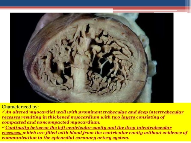
Noncompaction Cardiomyopathy

Cureus Left Ventricle Non Compaction Cardiomyopathy Admitted With Multiorgan Failure A Case Report
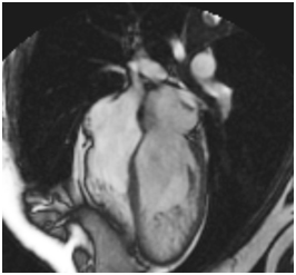
Left Ventricular Noncompaction

Incremental Value Of Contrast Echocardiography In The Diagnosis Of Left Ventricular Noncompaction
A Patient With Abnormalities Of The Coronary Arteries And Non Compaction Of The Left Ventricular Myocardium Resulting In Ischaemic Heart Disease Symptoms Dabek Folia Morphologica

Catastrophic Stroke In A Patient With Left Ventricular Non Compaction In Echo Research And Practice Volume 5 Issue 3 18

Wall Thickness Measurements In Lv Non Compaction Para Sternal Short Download Scientific Diagram
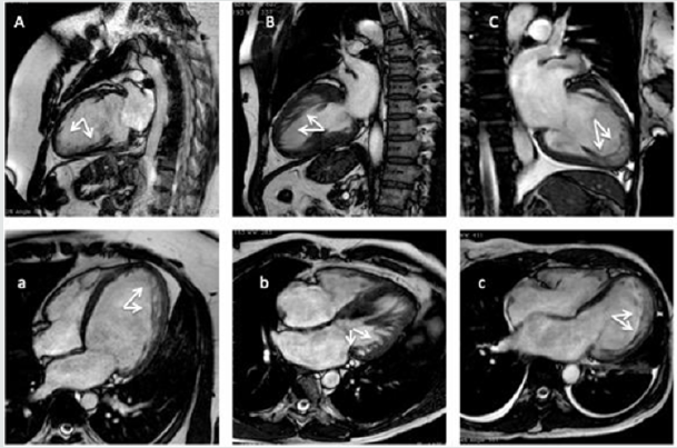
Non Compacted Cardiomyopathy Is There A Need Of A New Cardiomyopathy

Isolated Non Compaction Of Right Ventricular Myocardium A Rare Congenital Anomaly And Role Of Echocardiogram A Case Report

Left Ventricular Non Compaction Cardiomyopathy Cardiomyopathy Uk

The Different Clinical Scenario Of Left Ventricular Non Compaction Report Of Three Cases Oa Case Reports

Short Axis Echo View Of Lvnc

Assessment Of Left Ventricular Non Compaction In Adults Side By Side Comparison Of Cardiac Magnetic Resonance Imaging With Echocardiography Sciencedirect
Left Ventricular Non Compaction Cardiomyopathy Thoracic Key

Left Ventricular Noncompaction Intechopen

Left Ventricular Noncompaction

Ebstein S Anomaly With Left Ventricular Noncompaction And Bicuspid Aortic Valve Jacc Journal Of The American College Of Cardiology

Multiple Thrombi In The Lv In Non Compaction Cardiomyopathy American College Of Cardiology

Echocardiogram Lv Non Compaction Youtube
Echocardiography In The Diagnosis Left Ventricular Noncompaction Cardiovascular Ultrasound Full Text

Masking And Unmasking Of Isolated Noncompaction Of The Left Ventricle With Real Time Contrast Echocardiography Circulation Cardiovascular Imaging
Left Ventricular Non Compaction Lvnc

Affected Lv Walls In Lv Non Compaction Parasternal Short Axis Views At Download Scientific Diagram
Left Ventricular Non Compaction A Review Of Literature On Clinical Status And Meta Analysis Of Diagnostic And Clinical Management Methods
Http Www Ijcem Com Files Ijcem Pdf

Isolated Left Ventricular Noncompaction In Sub Saharan Africa A Clinical And Echocardiographic Perspective Circulation Cardiovascular Imaging

Left Ventricular Hypertrabeculation A Clinical Enigma Bmj Case Reports

Non Compaction Of The Left Ventricle Radiology Reference Article Radiopaedia Org

Left Ventricular Noncompaction Cardiomyopathy Lvnc Youtube

A Case Of Isolated Left Ventricular Noncompaction With Basal Ecg Tracing Strongly Suggestive For Type 2 Brugada Syndrome
Left Ventricular Non Compaction A Review Of Literature On Clinical Status And Meta Analysis Of Diagnostic And Clinical Management Methods

Left Ventricular Trabeculations In Athletes American College Of Cardiology
Left Ventricular Non Compaction A Review Of Literature On Clinical Status And Meta Analysis Of Diagnostic And Clinical Management Methods

Noncompaction Cardiomyopathy Case Presentation With Cardiac Magnetic Resonance Imaging Findings And Literature Review

Color Doppler In Lv Non Compaction Apical Four Chamber Views With Download Scientific Diagram

Isolated Left Ventricular Noncompaction Cardiomyopathy A Transient Disease

Left Ventricular Noncompaction Cardiomyopathy Circulation

Catastrophic Stroke In A Patient With Left Ventricular Non Compaction In Echo Research And Practice Volume 5 Issue 3 18

Hypertrophic Cardiomyopathy Associated With Left Ventricular Noncompaction Cardiomyopathy And Coronary Fistulae A Case Report One Genotype Three Phenotypes Revista Portuguesa De Cardiologia English Edition

Non Compaction Cardiomyopathy Heart
Www Onlinejacc Org Content 64 17 1840 Full Pdf

British Cardiovascular Society
Left Ventricular Noncompaction Cardiomyopathy In Pediatric Patients A Case Series Of A Clinically Heterogeneous Disease Springerlink

Contrast And 3d Echocardiography In Lv Non Compaction Apical Four Download Scientific Diagram
Q Tbn 3aand9gcrkapcl4baeka9fkrwrvwzj0ju90acr2gmm Sddxkhbkugmdsiw Usqp Cau
Onlinelibrary Wiley Com Doi Pdf 10 7863 Jum 12 31 10 1551

Limitations In The Diagnosis Of Noncompaction Cardiomyopathy By Echocardiography

Left Ventricular Trabeculations In Athletes American College Of Cardiology
Expanding The Impact Of Contrast Echocardiography
Q Tbn 3aand9gcqjvpxgcpbnml9epyktyhecskzgai Bug6daksbdocvsnqcknf Usqp Cau
Left Ventricular Non Compaction

Comparison Of Regional Systolic Myocardial Velocities In Patients With Isolated Left Ventricular Noncompaction And Patients With Idiopathic Dilated Cardiomyopathy Journal Of The American Society Of Echocardiography

Sotos Syndrome Isolated Left Ventricular Non Compaction Cardiomyopathy And Ventricular Pre Excitation A Case Report Sotos Syndrome Isolated Left Ventricular Non Compaction Cardiomyopathy And Ventricular Pre Excitation A Case Report

Isolated Left Ventricular Non Compaction Cardiomyopathy In Adults Journal Of Cardiology
Non Compaction Of The Left Ventricle Radiology Reference Article Radiopaedia Org

Figure 7 From Left Ventricular Noncompaction Imaging Findings And Diagnostic Criteria Semantic Scholar

Pedi Cardiology Lv Non Compaction
Role Of Cardiac Imaging Echocardiography Springerlink
Http Medreviews Com Sites Default Files 17 02 Ricm153 8 Min Pdf
Www Asecho Org Wp Content Uploads 18 03 Umland Case Studies Left Ventricular Noncompaction Pdf

Contrast Echo Lv Non Compaction Echocardiography Cardiac Ultrasound Youtube

Left Ventricular Non Compaction Jacc Journal Of The American College Of Cardiology

A Rare Case Of Biventricular Non Compaction Bmj Case Reports
Www Onlinejacc Org Content 64 17 1840 Full Pdf

Lv Non Compaction 2d Youtube

Reproducibility Of Echocardiographic Diagnosis Of Left Ventricular Noncompaction Sciencedirect
Www Asecho Org Wp Content Uploads 18 03 Umland Case Studies Left Ventricular Noncompaction Pdf

Improvement In Systolic Function In Left Ventricular Non Compaction Cardiomyopathy A Case Report Sciencedirect
Multimodality Imaging Of Left Ventricular Non Compaction Cardiomyopathy Associated With Situs Inversus

Thickness And Ratio Of Noncompacted And Compacted Layers Of The Left Ventricular Myocardium Evaluated In 56 Normal Fetuses By Two Dimensional Echocardiography
Noncompaction

Clinical And Echocardiography Features Of Diagnosed In Adulthood Isolated Left Ventricular Noncompaction A Case Series Study Huang Wh Sung Kt Tsai Jp Lo Ci Hsiao Cc Kuo Jy Su Ch Chen Mr

Non Compaction Cardiomyopathy Heart

Improvement In Systolic Function In Left Ventricular Non Compaction Cardiomyopathy A Case Report Sciencedirect

Noncompaction Cardiomyopathy Case Presentation With Cardiac Magnetic Resonance Imaging Findings And Literature Review Saeedan Mb Fathala Al Mohammed Tlh Heart Views
British Cardiovascular Society
Left Ventricular Noncompaction
Isolated Ventricular Non Compaction Syndrome Rare Cause Of Recurrent Stroke In Young A Case Report Insight Medical Publishing
Q Tbn 3aand9gcsudvexapv1fpwyogwseapqrfe8dmgm4o7b Nsgqplfuizcxwt Usqp Cau
Q Tbn 3aand9gctgvbjhjzmvzgqtpdqugi0pfi2jttwcg9oim77pctd0oyxhq5ib Usqp Cau

Apical Hypertrophic Cardiomyopathy Or Left Ventricular Non Compaction Thoracic Key
Expanding The Impact Of Contrast Echocardiography

Figure 1 From Images In Cardiovascular Medicine Overlapping Phenotypes Left Ventricular Noncompaction And Hypertrophic Cardiomyopathy Semantic Scholar

Incremental Value Of Contrast Echocardiography In The Diagnosis Of Left Ventricular Noncompaction
Frontiers Different Manifestations In Familial Isolated Left Ventricular Non Compaction Two Case Reports And Literature Review Pediatrics


