Non Compaction Cardiomyopathy Echo Criteria
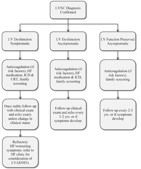
Presenting with Left Ventricular Non Compaction (LVNC, Group 1) or Idiopathic Dilated Cardiomyopathy (DCM, Group 2) At distance of an acute heart failure thrust (> 1 month) Newly diagnosed (less than 6 months) Diagnosis confirmed by echocardiography associated or not with a Magnetic Resonance Imaging (MRI) confirmed after central review.
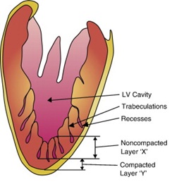
Non compaction cardiomyopathy echo criteria. The distinct morphologic features of LVNC cardiomyopathy can be readily identified by echocardiography;. An uninformative test has a sensitivity and specificity of 50%. J Am Soc Echocardiogr 12;.
The echocardiographic diagnosis of left ventricular non-compaction is difficult, and experienced readers disagree frequently. Left ventricular noncompaction (LVNC) cardiomyopathy is characterized by prominent myocardial trabeculations and deep recesses. Careful attention to suggested criteria and the use of other imaging modalities in difficult cases resolves most diagnostic disagreements.
Non-compaction cardiomyopathy, is a rare congenital cardiomyopathy that affects both children and adults. To determine clear cut echocardiographic criteria for isolated ventricular non-compaction (IVNC), a cardiomyopathy as yet "unclassified" by the World Health Organization. A trabeculated left ventricular mass above % of total mass with a sensitivity of 91.6% and a specificity of 86.5% is predictive of LVNC.
A novel echocardiographic criterion for non-compaction cardiomyopathy. 4 In 1932, Bellet and Gouley described the first case of noncompaction in an autopsy of a newborn infant with aortic atresia and coronary– ventricular fistula.8 LVNC without other cardiac abnormalities (isolated noncompaction cardiomyopathy). Current guidelines from professional organizations recommend different stra-tegies for diagnosing and treating patients with LVNC.
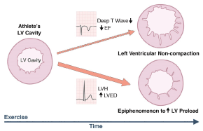
Left Ventricular Non-Compaction Cardiomyopathy:. Isolated noncompaction of left ventricular myocardium. 30 One hypothesis is that chronic increases in preload may be associated with.
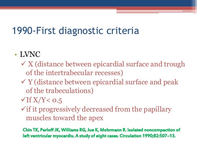
In the MOGE(S) classification endorsed by the World Heart Federation, cardiomyopathy is categorized by the following characteristics:. The following are key points to remember from this report on a multicenter retrospective study from the Netherlands that analyzed patients with noncompaction cardiomyopathy (NCCM). Chin TK, Perloff JK, Williams RG, et al.
There are currently no official diagnostic criteria for LVNC. Making the appropriate diagnosis using one of the well-defined echocardiographic diagnostic criteria requires adherence to a compulsory imaging protocol. During development, the majority of the heart muscle is a sponge-like meshwork of interwoven myocardial fibers.
To discuss diagnostic criteria for and the advantages and limitations of these imaging techniques;. The definition and classification of cardiomyopathy have evolved considerably in recent years. Whether left ventricular noncompaction (LVNC) is a distinct cardiomyopathy or a morphologic trait shared by different cardiomyopathies remains controversial.
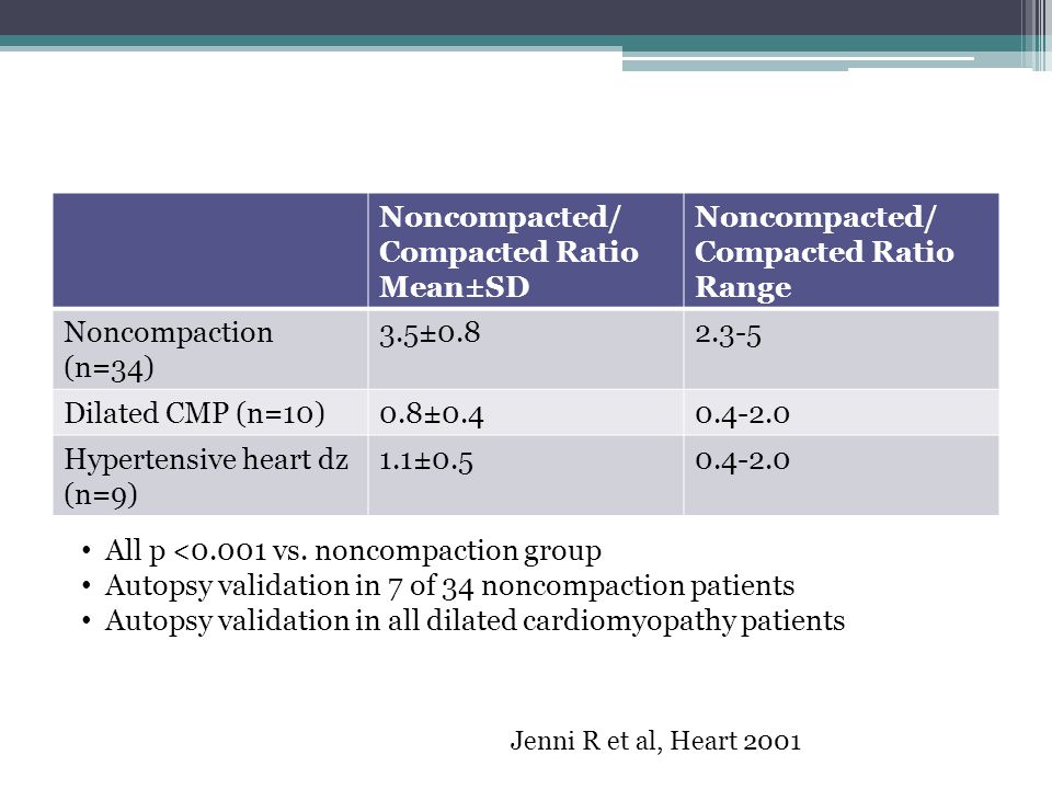
Nucifora G, Aquaro GD, Pingitore A, Masci PG, Lombardi M (10) Myocardial fibrosis in isolated left ventricular non-compaction and its relation to disease severity. As classification serves to bridge the gap between ignorance and knowledge the aim of the present study was to define clear cut echocardiographic diagnostic criteria for isolated ventricular non-compaction (IVNC) validated by necropsy.1 2 Although IVNC has been known for more than a decade. What is left ventricular non-compaction (LVNC)?.
Diagnostic Criteria Diagnosis can be made by echocardiography. Eur J Heart Fail 13(2):. (7) described another method to diagnose this entity:.
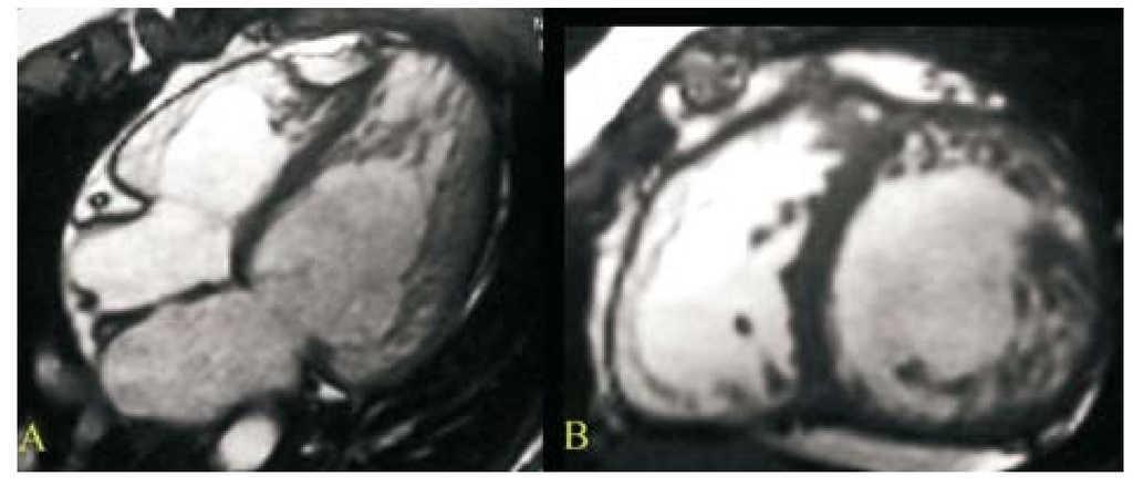
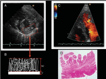
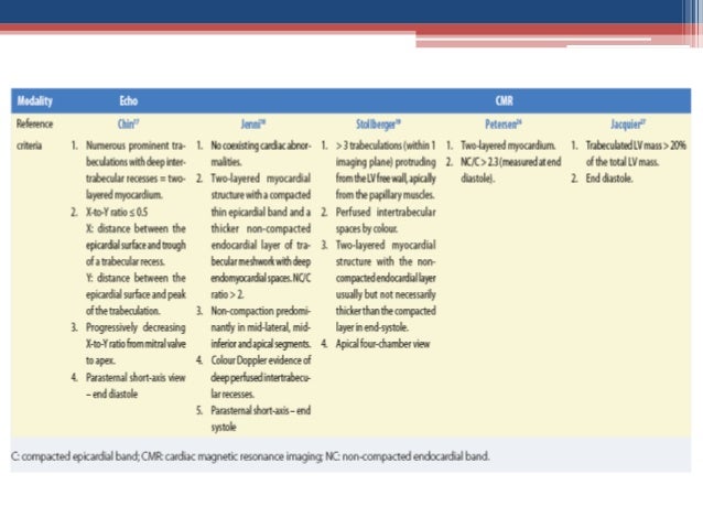
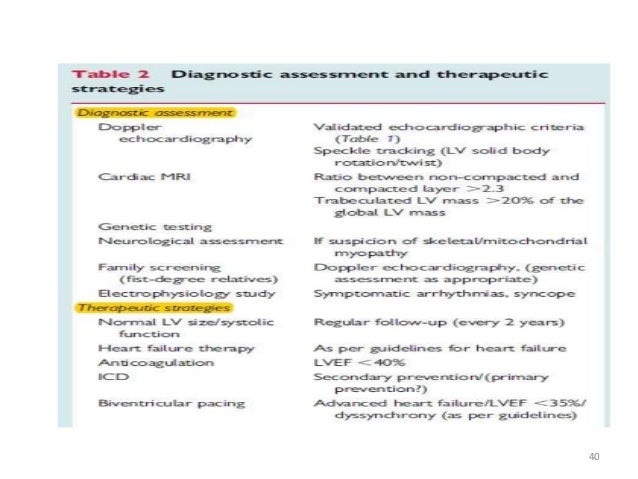
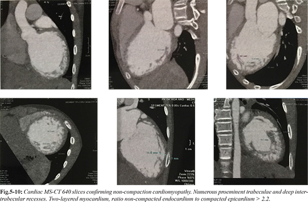
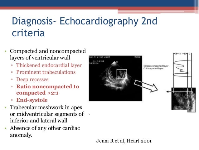
The ratio of noncompacted myocardium to compacted myocardium must be greater than 2.3 during the diastole (sensitivity of 86% and specificity of 99%). CMR and pathology findings. Echocardiographic diagnostic criteria have been proposed for the isolated form of LVNC and include (1) absence of coexisting cardiac abnormalities, (2) noncompaction to compaction ratio of ≥2:1 at end systole, (3) segmental thickening of the LV myocardium with a thin compacted epicardial layer and a thick noncompacted endocardial layer with.
Also called insulated non compaction of the ventricular myocardium (INVM), it is a rare form of congenital heart disease in which the tissue of the ventricular myocardium is not well constructed in terms of texture. Diagnostic criteria used in non-compaction cardiomyopathy. Left ventricular non-compaction (LV NC) is characterized by abnormal trabeculations that are mainly at the LV apex.
In very low pre-test probabilities consistent with the reported left ventricular noncompaction (LVNC) prevalence of 0.014% to 0.5%, neither cardiac magnetic resonance (CMR) criteria are very informative. There are five types of cardiomyopathy that are each recognized by echocardiography. Non-compaction cardiomyopathy (NCM) is a myocardial disorder, which is thought to occur due to the failure of left ventricle (LV) compaction during embryogenesis, leading to distinct morphological.
In 08, the European Society of Cardiology Working Group on Myocardial and Pericardial Diseases categorized LVNC. It can be associated with left ventricular dilation or hypertrophy, systolic or diastolic dysfunction, or both, or various forms of congenital heart disease. Current echocardiographic criteria for diagnosis typically include the following three:.
Interobserver agreement of the echocardiographic. Symptoms, Diagnosis and Treatment for LVNC. Jenni criteria (Heart 07).
This process is particularly apparent in the ventricles, and particu. Stöllberger C, et al. • The best diagnostic performance can be achieved if combined MRI criteria for the diagnosis are used.
Clinical guidelines help to support evidence-based practice in echocardiography, and all guidelines can be found in the ECHO journal. Left Ventricular Non-Compaction Case Studies Matt Umland, ACS, RDCS, FASE Aurora Health Care Milwaukee, WI Left Ventricular Noncompaction Cardiomyopathy • 1926 Grant - Malformed heart of a child • 1975 Dusek - Spongy Myocardium • 1984 Englberding – Echo Diagnosis of Myocardial Sinusoids • 1986 Jenni – Biventricular Sinusoids. - Presence of multiple echocardiographic trabeculations, particularly in the apex and free.
Similarly, our protocols help establish step-by-step procedures for performing roles within the echocardiography field, offering an advanced resource that can be beneficial across the board. Hence, echocardiography is the current gold standard for diagnosis of this entity. Stress echocardiography and left ventricular contractile reserve.
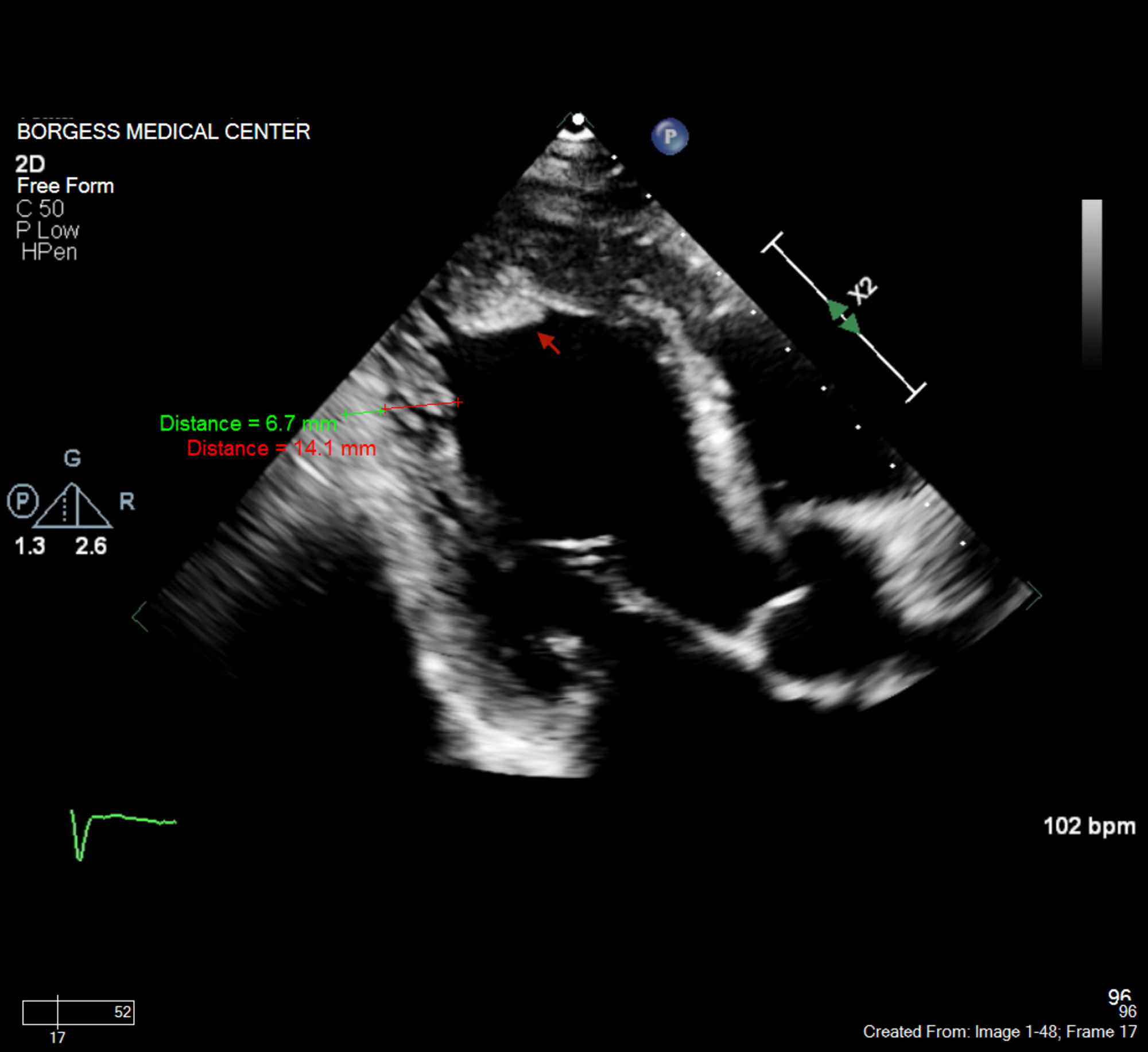
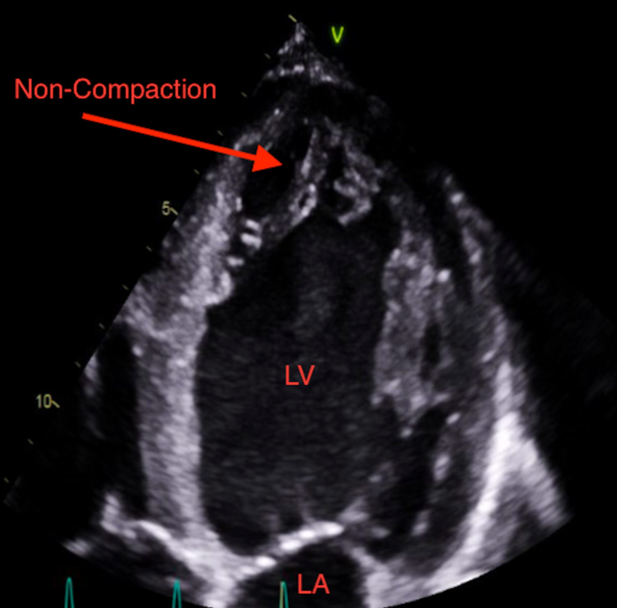
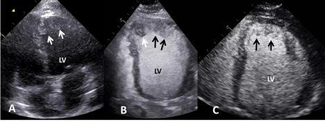
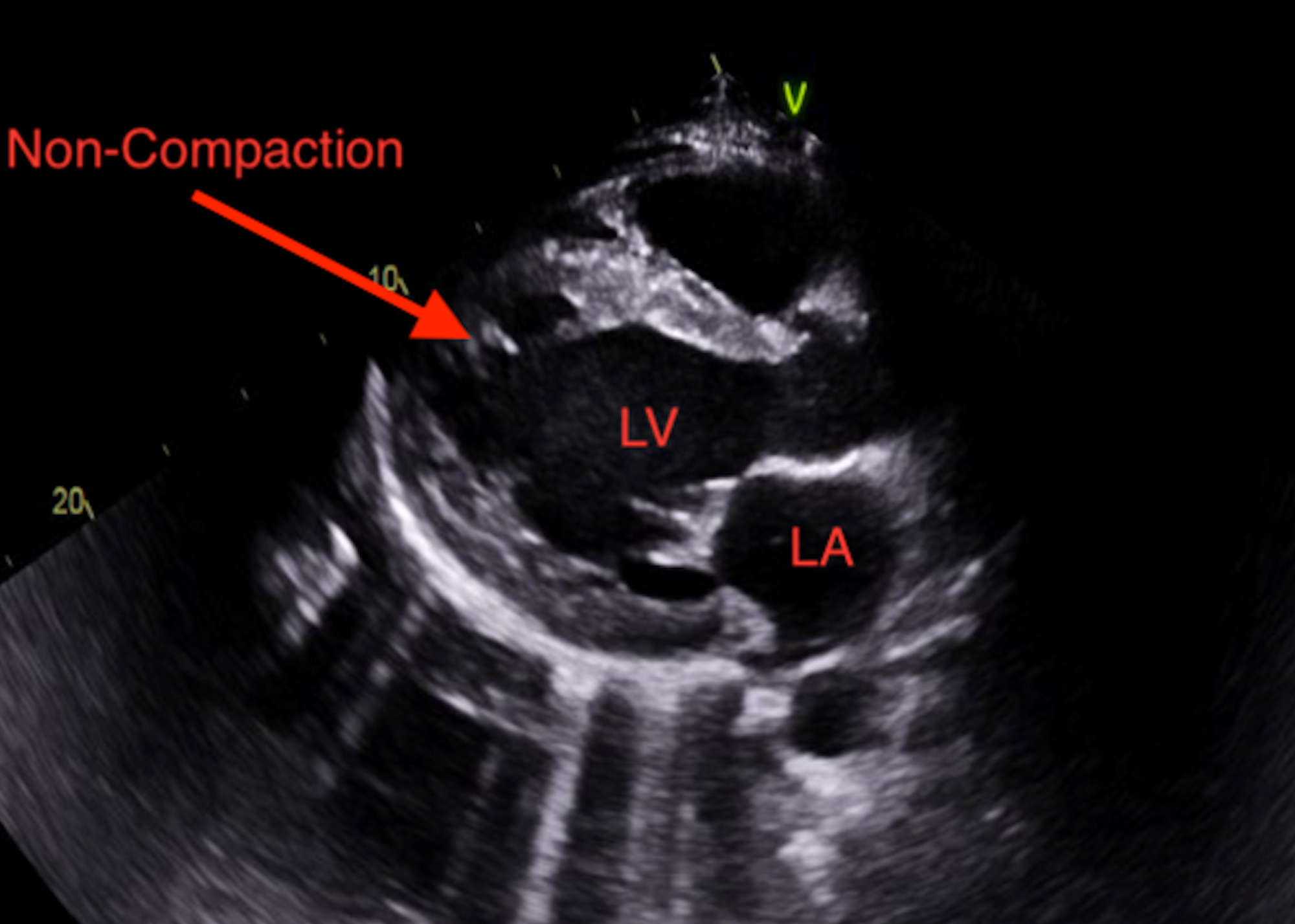
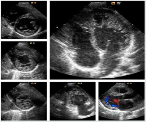
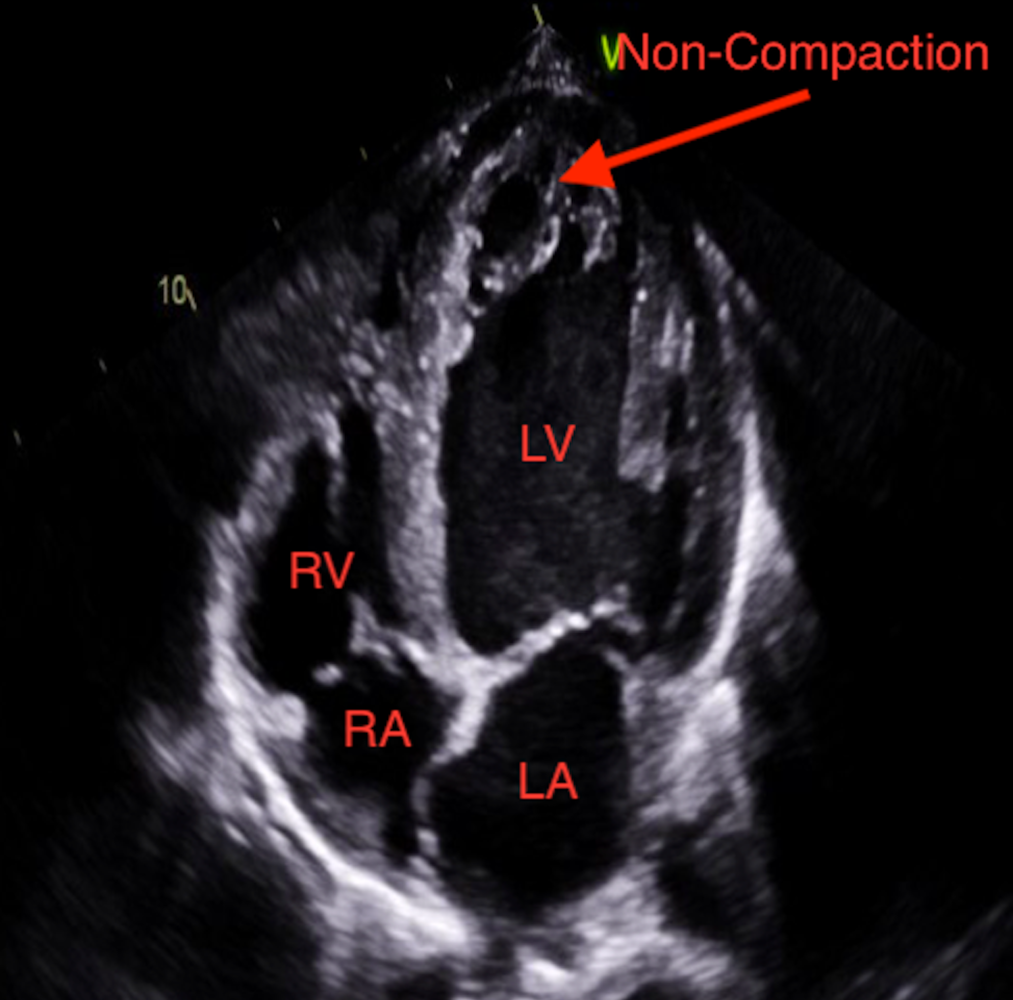
Left ventricular noncompaction is a rare unclassified cardiomyopathy with markedly prominent apical trabeculae with deep intertrabecular recesses (Fig. Non-compaction cardiomyopathy (NCCMP) LV wall has a spongy appearance. In left ventricular non-compaction cardiomyopathy (LVNC) the lower left chamber of the heart, called the left ventricle, contains bundles or pieces of muscle that extend into the chamber.
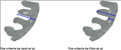
Later, Jacquier et al. Diagnosis- Echocardiography 2nd criteria • Compacted and noncompacted layers of ventricular wall Thickened endocardial layer Prominent trabeculations Deep recesses Ratio noncompacted to compacted >2:1 End-systole • Trabecular meshwork in apex or midventricular segments of inferior and lateral wall • Absence of any other cardiac anomaly. Non-compaction cardiomyopathy is a rare cardiac disorder which commonly goes undiagnosed until post-mortem, although diagnosis through echocardiogram, CT, or MRI is possible and there is criterion.
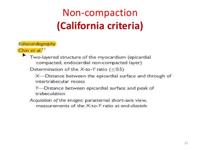
The Jenni criteria, which stress the presence of a 2-layered structure, and the Chin criteria, which focus on the depth of the recess compared with the height of the trabecula 8, 9. In both sets, it is important that there are no other cardiac structural abnormalities, such as semilunar valve obstruction or coronary artery anomalies. 1 Left ventricular non-compaction was classified as a primary cardiomyopathy by the American Heart Association in 06, 2 however, remains unclassified by the European.
Criteria (e.g., the compacted to noncompacted ratio) should differ by age at diagnosis. As normal development progresses, these trabeculated structures undergo significant compaction that transforms them from spongy to solid. Chin et al 2.
1, communication with the intertrabecular space demonstrated by Doppler, absence of coexisting cardiac abnormalities, and presence of multiple prominent trabeculations in end-systole 22. Distinction between LV NC and non-specific dilated cardiomyopathies (DCMs) remains often challenging. There are frequent doubtful cases, that need multimodality confirmation.
In seven out of a series of 34 patients with IVNC the in vivo echocardiographic characteristics were validated against the anatomical. Individual variability is extreme, and trabeculae represent a sort of individual “cardioprinting.” By itself, the diagnosis of LVNC does not coincide with that of a “cardiomyopathy” because it. Left ventricular noncompaction (LVNC) describes a ventricular wall anatomy characterized by prominent left ventricular (LV) trabeculae, a thin compacted layer, and deep intertrabecular recesses.
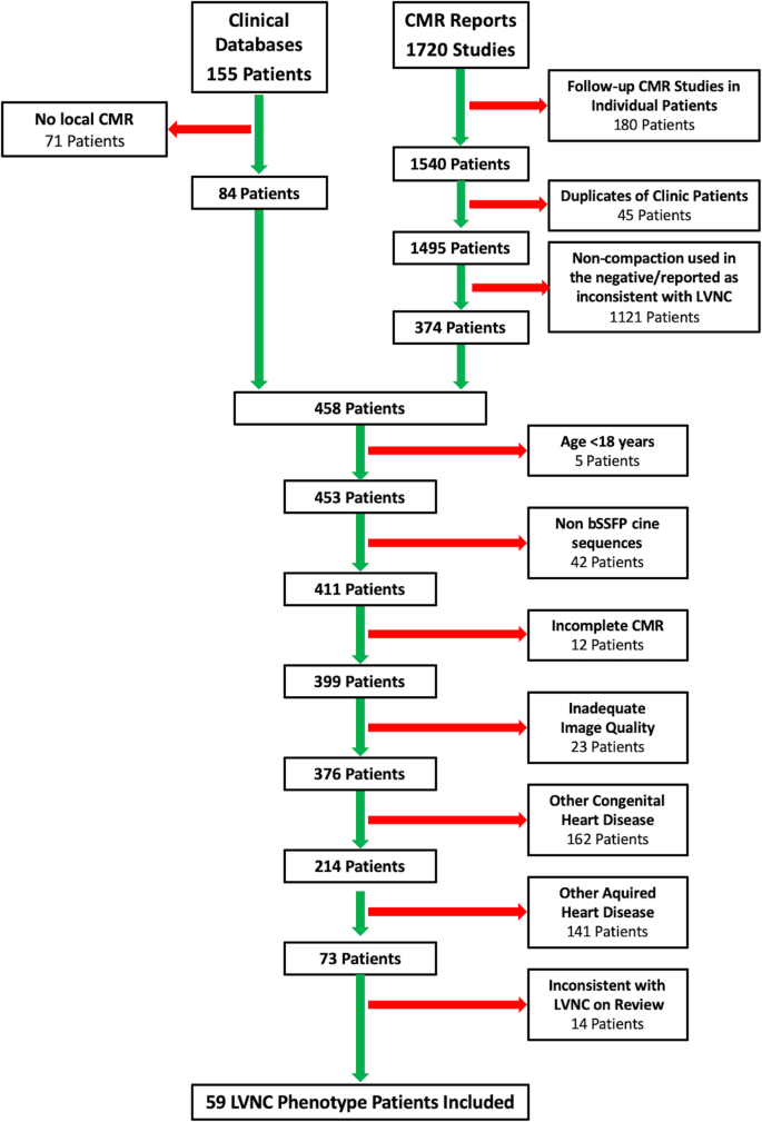
We sought to find additive tools comparing the longitudinal strain characteristics …. Criteria for diagnosis by CMR:. The objectives of this article are to review the imaging findings of left ventricular noncompaction (LVNC) at echocardiography, cardiac MRI, and MDCT;.
Left ventricular noncompaction (LVNC) is a relatively new entity. Cardiomyopathy can be separated into primary (genetic, mixed, or acquired) and secondary categories. • Cardiac magnetic resonance imaging can reliably diagnose left ventricular non-compaction cardiomyopathy.
Pujadas S, Bordes R, Bayes-Genis A (05) Ventricular non-compaction cardiomyopathy:. These criteria include a bilayered myocardium, a noncompacted to compacted ratio >2 :. Left ventricular non-compaction (LVNC) is a condition of the heart where the walls of the left ventricle (the bottom chamber of the left side of the heart) are non-compacted, causing channels to form in the heart muscle.
To get enough information for the diagnosis, your physician might require various tests and information. (6) described the criteria for the diagnosis by CMR:. By Michael Crawford, MD, Editor SYNOPSIS:.
Left ventricular noncompaction (LVNC) is a cardiomyopathy associated with sporadic or familial disease, the latter having an autosomal dominant mode of transmission.Echocardiography is the current gold standard for diagnosis of this entity, but with the risk of over diagnosis and under diagnosis. Reduced left ventricular compacta thickness:. During development, the heart muscle is a sponge-like network of muscle fibers.
Stress echocardiography is a useful tool in guiding management in DCM, by identifying the presence or absence of contractile reserve (improvement in wall motion score, fractional shortening, or EF) during dobutamine infusion (10–40 mcg/kg/min). It is characterized by trabeculated myocardium with adjacent deep intertrabecular recesses communicating with the LV cavity .Prominent myocardial trabeculations were first identified in a variety of congenital heart defects and then in the absence of any other structural heart disease 2, 3. These pieces of muscles are called trabeculations.
Jenni et al 3. Ratio of X*/Y† <0.5. This causes channels to form in the heart muscle, called trabeculations.
• Differentiation of LVNC from other cardiomyopathies and normal hearts is possible. There are 2 sets of echocardiographic criteria for IVNC diagnosis:. The disease is not widely known and its diagnosis mostly missed.
Gebhard C, Stähli BE, Greutmann M, et al. This gives the left ventricle a 'spongy' look (a bit like honeycomb). NCCM is characterized by excessive trabeculations typically involving the left ventricle (LV) with >2:1 ratio of noncompacted:compacted myocardium.
Gati et al alluded to the overreporting of noncompaction in healthy athletes, with 8.1% of African/Afro-Caribbean males fulfilling diagnostic criteria by echocardiogram for noncompaction cardiomyopathy, but having no real adverse events when followed longitudinally. J Am Coll Cardiol 18;71:711-722. Non-compaction of the left ventricle, also known as spongiform cardiomyopathy or left ventricular non-compaction (LVNC) is a phenotype of hypertrophic ventricular trabeculations and deep interventricular recesses.It has been hypothesized to result from arrest of normal myocardial compaction during embryogenesis, although acquired cases have also been reported.
3 The clinical diagnosis is predominantly reliant on three. And to describe pitfalls that can lead to misinterpretation of findings of LVNC. Left ventricular non-compaction, also known as LVNC, spongy myocardium or hypertrabeculation syndrome, is a pathologic cardiac condition in which the myocytes exhibit a “spongy” appearance.
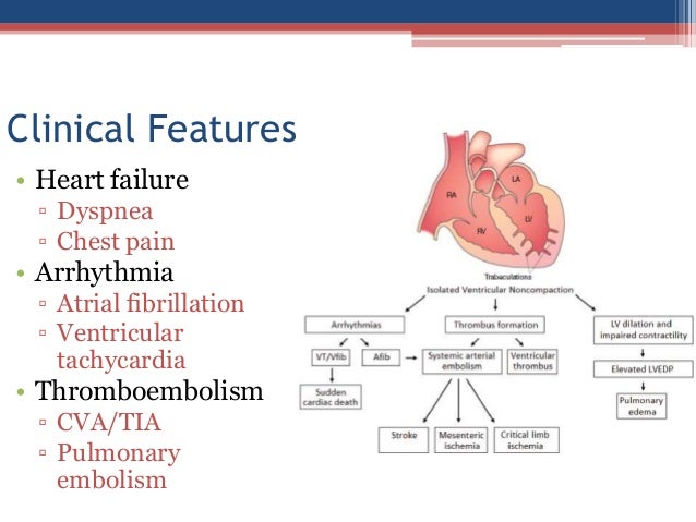
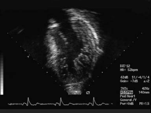
Studies in heart failure patients demonstrate a high prevalence of myocardial trabeculations, raising the potential diagnosis of LVNC. These are best visualized on color flow Doppler of the left ventricle using apical windows. 1-3 The clinical spectrum of the disorder ranges from being completely asymptomatic to progressive left ventricular (LV) systolic impairment, a tendency to fatal arrhythmias and systemic thromboembolic events.
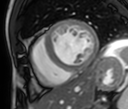
Noncompaction cardiomyopathy used to be called “spongy myocardium” due to its spongy appearance. Morphofunctional (M), organ involvement (O), genetic or familial inheritance (G), etiological annotation (E), and stage (S) 3. Left ventricular non-compaction (LVNC) is described as a distinct cardiomyopathy characterized by the presence of a bilayered myocardium with prominent trabeculations (Figure 1).
Left ventricular non-compaction, the most recently classified form of cardiomyopathy, is characterised by abnormal trabeculations in the left ventricle, most frequently at the apex. Left ventricular noncompaction (LVNC) cardiomyopathy is morphologically characterized by prominent myocardial trabeculations and deep recesses. The precise stage of development and the natural history of the disorder are not fully understood.
A study of eight cases. Evaluates the trabeculations at the left ventricle (LV) apex, using the short axis and apical views and the free wall, at end-diastole. Our echocardiography laboratory was consulted to determine whether a patient's echocardiogram would fulfill the criteria of left ventricular noncompaction cardiomyopathy (LVNC).
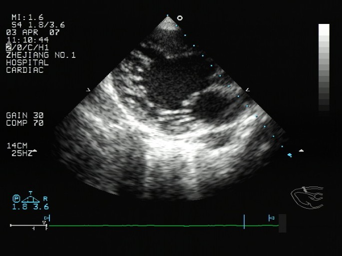
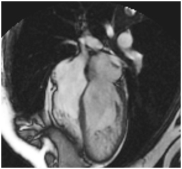
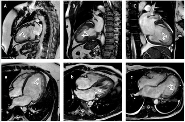
Images of the left ventricle showed a 2-layer structure with a compacted, thin epicardial band and a much thicker noncompacted endocardial layer of trabecular meshwork. LVNC is a condition of the heart where the walls of the left ventricle (the bottom chamber of the left side of the heart) are non-compacted. Hypertrophic Cardiomyopathy Echocardiographic Diagnosis Left Ventricular Hypertrophy 15 mm (Asymmetric >> Symmetric) In the absence of another cardiovascular or systemic disease associated with LVH or myocardial wall thickening Gersh, BJ, et al.
Affected individuals are at risk of left or right.

Isolated Left Ventricular Noncompaction In Sub Saharan Africa A Clinical And Echocardiographic Perspective Circulation Cardiovascular Imaging

Incremental Value Of Contrast Echocardiography In The Diagnosis Of Left Ventricular Noncompaction
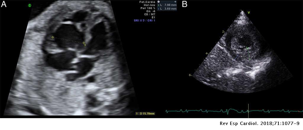
Undulating Clinical Course Of Noncompaction Cardiomyopathy Revista Espanola De Cardiologia English Edition
Non Compaction Cardiomyopathy Echo Criteria のギャラリー

Diagnostic Assessment And Therapeutic Strategies Download Table
Noncompaction Cardiomyopathy

Non Compaction Cardiomyopathy Heart

Left Ventricular Noncompaction A Diagnostically Challenging Cardiomyopathy Semantic Scholar
Echocardiography In The Diagnosis Left Ventricular Noncompaction Cardiovascular Ultrasound Full Text

British Cardiovascular Society

Catastrophic Stroke In A Patient With Left Ventricular Non Compaction In Echo Research And Practice Volume 5 Issue 3 18
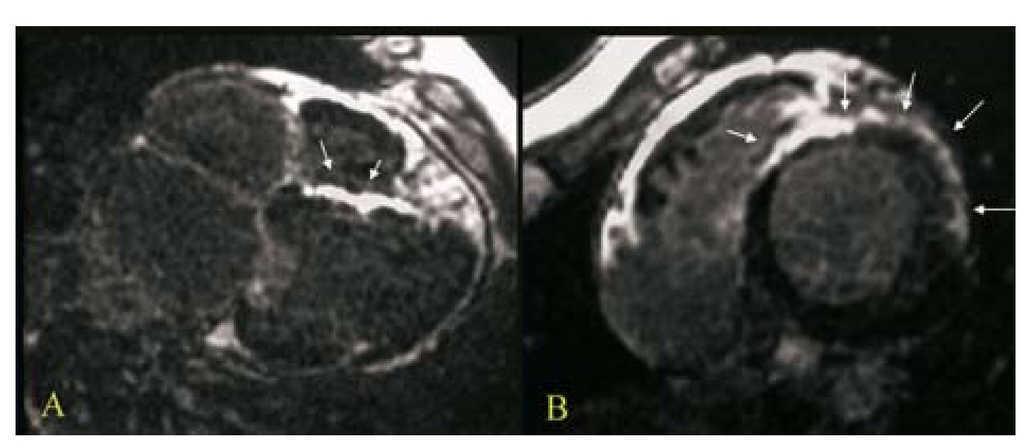
Late Gadolinium Enhancement In Non Compaction Cardiomyopathy Revista Espanola De Cardiologia English Edition
Cureus A Case Of Left Ventricular Noncompaction Presenting As Atrial Fibrillation
Www Asecho Org Wp Content Uploads 18 03 Umland Case Studies Left Ventricular Noncompaction Pdf
Left Ventricular Non Compaction A Review Of Literature On Clinical Status And Meta Analysis Of Diagnostic And Clinical Management Methods
Www Internationaljournalofcardiology Com Article S0167 5273 09 4 Pdf

Echocardiography Fails To Detect Left Ventricular Noncompaction In A Cohort Of Patients With Noncompaction On Cardiac Magnetic Resonance Imaging Diwadkar 17 Clinical Cardiology Wiley Online Library
Cureus Heart Failure Secondary To Left Ventricular Non Compaction Cardiomyopathy In A 26 Year Old Male

Isolated Left Ventricular Noncompaction Report Of A Case From Ibadan Nigeria Ogah Os Adebayo O Aje A Koya Fk Towoju O Adesina Jo Adeoye Am Adebiyi Oladapo Oo Falase Ao

Left Ventricular Noncompaction Jacc Journal Of The American College Of Cardiology
Late Gadolinium Enhancement In Non Compaction Cardiomyopathy Revista Espanola De Cardiologia English Edition

Left Ventricular Non Compaction Cardiomyopathy Cardiomyopathy Uk

Isolated Form Of Left Ventricular Non Compaction Cardiomyopathy Lvnc As A Rare Cause Of Heart Failure Intechopen
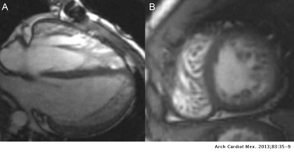
Left Ventricle Non Compaction Cardiomyopathy Different Clinical Scenarios And Magnetic Resonance Imaging Findings Archivos De Cardiologia De Mexico
Www Ecronicon Com Eccea Pdf Eccea 02 Pdf

Non Compaction Cardiomyopathy Heart

Clinical And Echocardiography Features Of Diagnosed In Adulthood Isolated Left Ventricular Noncompaction A Case Series Study Huang Wh Sung Kt Tsai Jp Lo Ci Hsiao Cc Kuo Jy Su Ch Chen Mr

Improvement In Systolic Function In Left Ventricular Non Compaction Cardiomyopathy A Case Report Sciencedirect
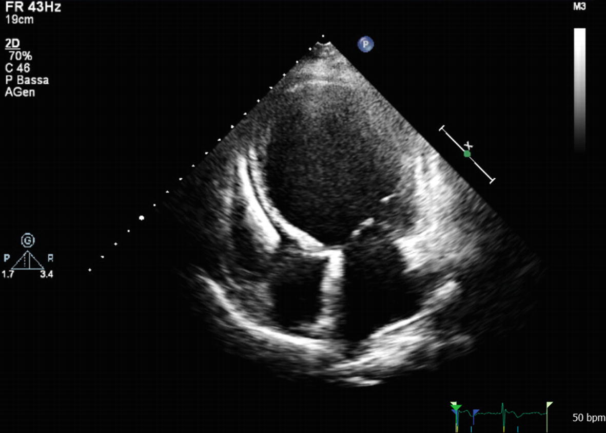
Role Of Cardiac Imaging Echocardiography Springerlink

Left Ventricular Noncompaction

Left Ventricular Noncompaction Jacc Journal Of The American College Of Cardiology
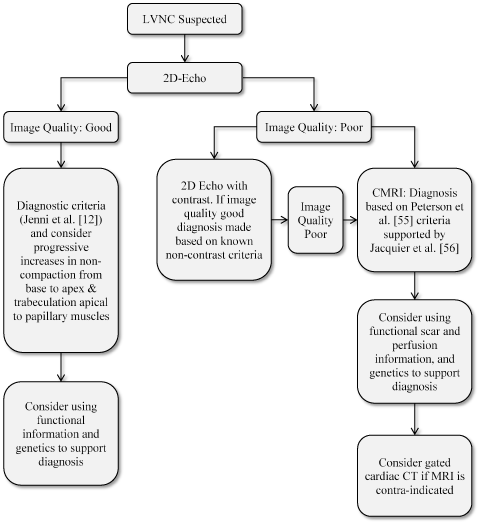
Left Ventricular Non Compaction A Review Of Literature On Clinical Status And Meta Analysis Of Diagnostic And Clinical Management Methods

Echocardiographic Comparison Between Left Ventricular Non Compaction And Hypertrophic Cardiomyopathy Sciencedirect

Two Potentially Fatal Surprises In The Preoperative Assessment Of An Asymptomatic Young Adult Revista Portuguesa De Cardiologia

Improvement In Systolic Function In Left Ventricular Non Compaction Cardiomyopathy A Case Report Sciencedirect

Non Compaction Of The Left Ventricle Radiology Reference Article Radiopaedia Org
Left Ventricular Non Compaction A Review Of Literature On Clinical Status And Meta Analysis Of Diagnostic And Clinical Management Methods
Noncompaction Cardiomyopathy

Color Doppler In Lv Non Compaction Apical Four Chamber Views With Download Scientific Diagram
Left Ventricular Non Compaction

Non Compaction Cardiomyopathy Heart

Reversible De Novo Left Ventricular Trabeculations In Pregnant Women Circulation

Myocardial Deformation Pattern In Left Ventricular Non Compaction Comparison With Dilated Cardiomyopathy Abstract Europe Pmc
Multimodality Imaging Of Left Ventricular Non Compaction Cardiomyopathy Associated With Situs Inversus
Q Tbn 3aand9gcrkapcl4baeka9fkrwrvwzj0ju90acr2gmm Sddxkhbkugmdsiw Usqp Cau

Reduced Left Ventricular Compacta Thickness A Novel Echocardiographic Criterion For Non Compaction Cardiomyopathy Thoracic Key

British Cardiovascular Society

Are We Getting Closer To Risk Stratification In Left Ventricular Noncompaction Cardiomyopathy Journal Of The American Heart Association
Left Ventricular Non Compaction A Review Of Literature On Clinical Status And Meta Analysis Of Diagnostic And Clinical Management Methods

Clinical And Echocardiography Features Of Diagnosed In Adulthood Isolated Left Ventricular Noncompaction A Case Series Study Huang Wh Sung Kt Tsai Jp Lo Ci Hsiao Cc Kuo Jy Su Ch Chen Mr

Isolated Non Compacted Left Ventricle A Diagnostic Dilemma Chaturvedi M Singh O Agarwal A Heart India

Noncompaction Cardiomyopathy Case Presentation With Cardiac Magnetic Resonance Imaging Findings And Literature Review Saeedan Mb Fathala Al Mohammed Tlh Heart Views
Left Ventricular Noncompaction

Masking And Unmasking Of Isolated Noncompaction Of The Left Ventricle With Real Time Contrast Echocardiography Circulation Cardiovascular Imaging
Non Compacted Cardiomyopathy Is There A Need Of A New Cardiomyopathy

Limitations In The Diagnosis Of Noncompaction Cardiomyopathy By Echocardiography

Isolated Left Ventricular Non Compaction Controversies In Diagnostic Criteria Adverse Outcomes And Management Heart

Proposed Diagnostics Criteria Of Left Ventricular Non Compaction Download Table
Noncompaction Cardiomyopathy
Q Tbn 3aand9gct7b9znw9upokpn6siznu0irwjuxzcoqrn66p Z7sr9z Czbo Usqp Cau

Wall Thickness Measurements In Lv Non Compaction Para Sternal Short Download Scientific Diagram

Left Ventricular Noncompaction

Echocardiography In The Diagnosis Left Ventricular Noncompaction Topic Of Research Paper In Clinical Medicine Download Scholarly Article Pdf And Read For Free On Cyberleninka Open Science Hub

Table 1 From Diagnosis Of Left Ventricular Non Compaction In Patients With Left Ventricular Systolic Dysfunction Time For A Reappraisal Of Diagnostic Criteria Semantic Scholar
Cardiovascular Magnetic Resonance Based Diagnosis Of Left Ventricular Non Compaction Cardiomyopathy Impact Of Cine Bssfp Strain Analysis Journal Of Cardiovascular Magnetic Resonance Full Text

Left Ventricular Trabeculations In Athletes American College Of Cardiology
Q Tbn 3aand9gcqmiymjbnajimtfkhbhjwdjrfdxd9uib D2ewnxynqm6 Nuojp8 Usqp Cau

Noncompaction Cardiomyopathy Case Presentation With Cardiac Magnetic Resonance Imaging Findings And Literature Review
Onlinelibrary Wiley Com Doi Pdf 10 1111 Echo
Non Compaction Of The Left Ventricle Radiology Reference Article Radiopaedia Org
Left Ventricular Noncompaction Cardiomyopathy Thoracic Key

Left Ventricular Noncompaction A Distinct Cardiomyopathy Or A Trait Shared By Different Cardiac Diseases Sciencedirect
Left Ventricular Non Compaction

Isolated Left Ventricular Noncompaction Cardiomyopathy A Transient Disease

Left Ventricular Noncompaction Cardiomyopathy Shemisa Cardiovascular Diagnosis And Therapy

Noncompaction Of The Left Ventricle A New Cardiomyopathy Is Presented To The Clinician

Isolated Left Ventricular Noncompaction Cardiomyopathy A Transient Disease

Left Ventricular Noncompaction Cardiomyopathy Shemisa Cardiovascular Diagnosis And Therapy

Isolated Left Ventricular Non Compaction Cardiomyopathy In Adults Journal Of Cardiology

Left Ventricular Non Compaction Cardiomyopathy The Lancet

Contrast And 3d Echocardiography In Lv Non Compaction Apical Four Download Scientific Diagram
1
Echocardiography Conference Connie Tsao Jan 21 Ppt Video Online Download

Left Ventricular Noncompaction Cardiomyopathy Shemisa Cardiovascular Diagnosis And Therapy

Left Ventricular Noncompaction Intechopen
Cureus Heart Failure Secondary To Left Ventricular Non Compaction Cardiomyopathy In A 26 Year Old Male

Left Ventricular Noncompaction Cardiomyopathy Lvnc Youtube

Left Ventricular Trabeculations In Athletes American College Of Cardiology

Incremental Value Of Contrast Echocardiography In The Diagnosis Of Left Ventricular Noncompaction
Non Compacted Cardiomyopathy Is There A Need Of A New Cardiomyopathy
Left Ventricular Non Compaction A Review Of Literature On Clinical Status And Meta Analysis Of Diagnostic And Clinical Management Methods
Cureus Heart Failure Secondary To Left Ventricular Non Compaction Cardiomyopathy In A 26 Year Old Male
Non Compaction Cardiomyopathy Misdiagnosed As Dilated Cardiomyopathy

Left Ventricle Non Compaction Cardiomyopathy Different Clinical Scenarios And Magnetic Resonance Imaging Findings Archivos De Cardiologia De Mexico
Www Journal Of Cardiology Com Article S0914 5087 14 4 Pdf
Www Asecho Org Wp Content Uploads 18 03 Umland Case Studies Left Ventricular Noncompaction Pdf
Noncompaction Cardiomyopathy

Adult Left Ventricular Noncompaction Jacc Cardiovascular Imaging

Adult Left Ventricular Noncompaction Reappraisal Of Current Diagnostic Imaging Modalities Sciencedirect

Echocardiogram Lv Non Compaction Youtube
Www Onlinejacc Org Content 64 17 1840 Full Pdf
Left Ventricular Noncompaction

Multiple Thrombi In The Lv In Non Compaction Cardiomyopathy American College Of Cardiology

Limitations In The Diagnosis Of Noncompaction Cardiomyopathy By Echocardiography

Isolated Left Ventricular Noncompaction In Sub Saharan Africa A Clinical And Echocardiographic Perspective Circulation Cardiovascular Imaging


